
Figure 2. Silibinin potentiates IR-induced G2/M arrest, inhibition in cell proliferation and augments the oxidative stress selectively in PCa cells.
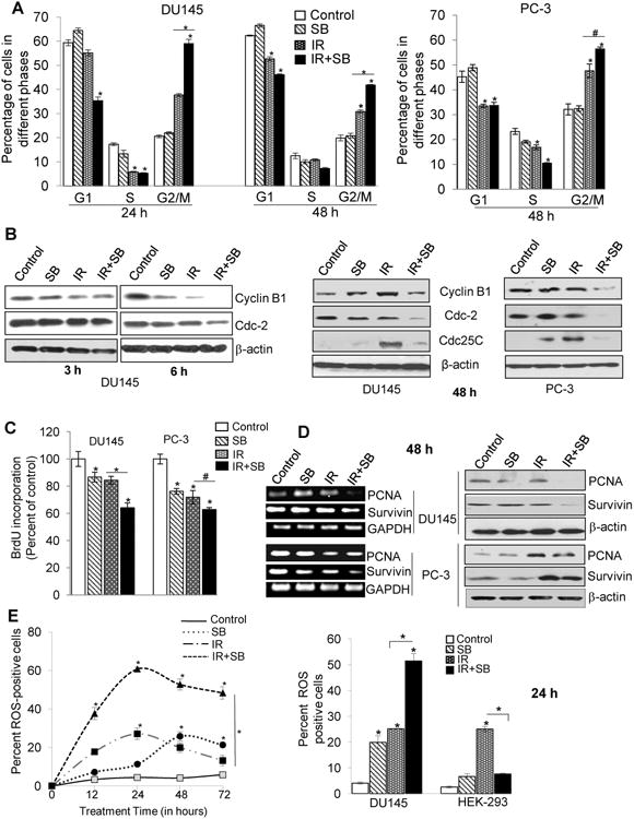
DU145 and PC-3 cells were exposed to ionizing radiation (IR) with or without silibinin (SB). After treatment time points, cells were processed for cell cycle analysis using saponin-PI staining (A) Quantitative data showing cell cycle distribution in DU145 (left panel) and PC-3 cells (right panel) after treatment with IR (5 Gy) and/or SB (25 μM). (B) Western blots for G2/M cell cycle related proteins at 3 h, 6 h and 48 h. Cell proliferation rate in cells was assessed by BrdU incorporation assay. (C) Percent BrdU incorporation was calculated with respect to control after 48 h treatment in both DU145 and PC-3 cells. (D) RT-PCR and immunoblotting analysis of PCNA and survivin proteins after 48 h treatment. (E) For oxidative stress analysis, Cells were analyzed for DCF fluorescence by flow cytometry, after treatment with IR (5 Gy) and/or SB (25 μM). Percent positive cells were those with a fluorescent intensity >102 on the histogram. Graph showing change in the DCF positive cells in different groups after 12-72 h of treatment of DU145 cells. Bar diagram showing DCF-positive cells for DU145 and HEK-293 cells after 24 h of treatment.