
Figure 3. Silibinin potentiates radiation-induced apoptosis and attenuates DNA repair activation signaling in PCa cells.
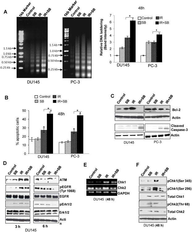
(A) Representative pictures (left panel) and quantitative data (right panel) showing DNA laddering in DU145 and PC-3 cells treated with IR (5 Gy) and/or silibinin (25 μM) for 48 h. (B) Graphical data depicting percent cells positive for apoptosis after Acridine orange –EtBr staining. (C) Western blot analysis of Bcl-2 and cleaved caspase-3 in DU145 and PC-3 cells after 48 h of treatment. (D) Immunoblotting for damage signaling molecules activated in response to IR (5 Gy) and/or SB (25 μM). (E) RT-PCR for cell cycle check-point regulators Chk1 and Chk2; and (F) phospho/total Chk1 (threonine 345 and serine 296) and Chk2 levels (threonine 68) in DU145 cells.