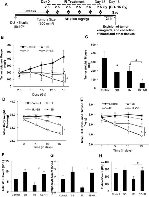
Figure 5. Silibinin treatment enhances radiation-induced tumor growth inhibition of human PCa DU145 xenograft in athymic nude mice.

(A) Diagrammatic representation of the time line followed for the tumor study. Mice were subcutaneously injected with DU145 cells (6×10⁶) mixed with Matrigel (1:1) and monitored for tumor growth till the tumor size reached ∼200 mm3. Then mice were treated with IR (2.5 Gy) with a gap of two days between two IR fractions, with or without SB (200 mg/kg), which was given 5 days/week. Control and IR alone group of mice were gavaged with 0.5% CMC in saline. The treatment was continued till the cumulative irradiation dose reached 15 Gy. Twenty four hours after the final fraction of IR (day 16), the tumors were excised and processed further for immune-histochemical staining. (B) Tumor volume/mouse as a function of cumulative radiation dose, (C) tumor weight/mouse at the end of study, (D) mean body weight/mouse, and (E) average diet consumption/mouse/day were analyzed as detailed in Materials and Methods. Data shown in mean ± SE from 8 mice in each group. Effect of IR and/or silibinin was also checked on the hematopoietic system at the end of the experiment (F) Mean WBC count/ mouse, (G) Mean lymphocyte count/mouse and (H) represents mean platelet count/mouse. P<0.05 ($); P<0.01 (#), P<0.001(*) compared with respective control.
