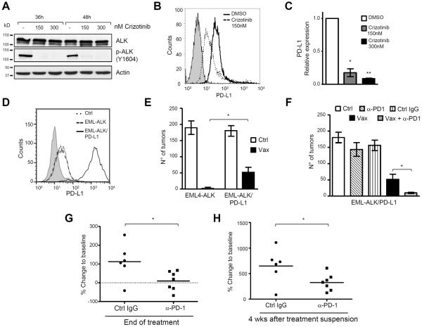
Figure 5. Blockade of the PD-1/PD-L1 pathway restores the efficacy of the ALK vaccine against cells expressing high PD-L1.
A, Western blot of H3122 cells treated with different crizotinib concentrations and collected at the indicated time points. Membranes were blotted with the indicated antibodies. B, PD-L1 protein expression was evaluated by flow cytometry in H3122 cells treated with 150nM crizotinib for 24 hours. C, PD-L1 mRNA expression was evaluated by qRT-PCR in crizotinib-treated cells. D, PD-L1 expression was evaluated by flow cytometry in ASB-XIV cells (Ctrl), EML4-ALK ASB-XIV (EML-ALK) and in EML4-ALK ASB-XIV transduced with PD-L1 (EML4-ALK/PD-L1). E, Mean tumor numbers in lungs from mice injected with the indicated ASB-XIV cells (n= 5 mice for each group). F, Mean tumor numbers in lungs from mice with the indicated treatments (n= 6–8 mice for each group). Data are represented as mean (±SEM). G and H, Quantification of volume changes compared to baseline tumors in ALK mice treated with control IgG (n=6 mice) or anti-PD-1 antibody (n=7 mice) at the end of treatment (G) and at 4 weeks after treatment suspension (H). Horizontal bars represent means. Data are from two independent experiments. *, P<0.05; **, P<0.005; ***, P<0.0005.

