Figure 3.
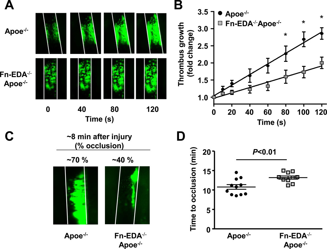
Fn-EDA−/−Apoe−/− mice are protected from experimental thrombosis of the carotid artery. A. Representative microphotographs of thrombus growth in FeCl3-injured carotid arteries as visualized by upright intravital microscopy. Platelets were labeled with calcein green. White lines delineate the arteries. B. The fold increase in diameter was calculated by dividing the diameter of the thrombus at time (n) by the diameter of the same thrombus at time (0) (defined as the time point at which the thrombus diameter first reached 100 µm). Slopes over time showed that the rate of thrombus growth in Fn-EDA−/−Apoe−/− mice (slope: 0.008 ± 0.001) was decreased when compared with Apoe−/− mice (slope: 0.016 ± 0.001). C. Representative microphotographs depicting percentage occlusion ~8 minutes after FeCl3-induced injury. Platelets were labeled with calcein green. White lines delineate the arteries D. Mean time to complete occlusion of FeCl3injured carotid artery. Data are presented as mean ± SEM. N=9–10 mice/group.

