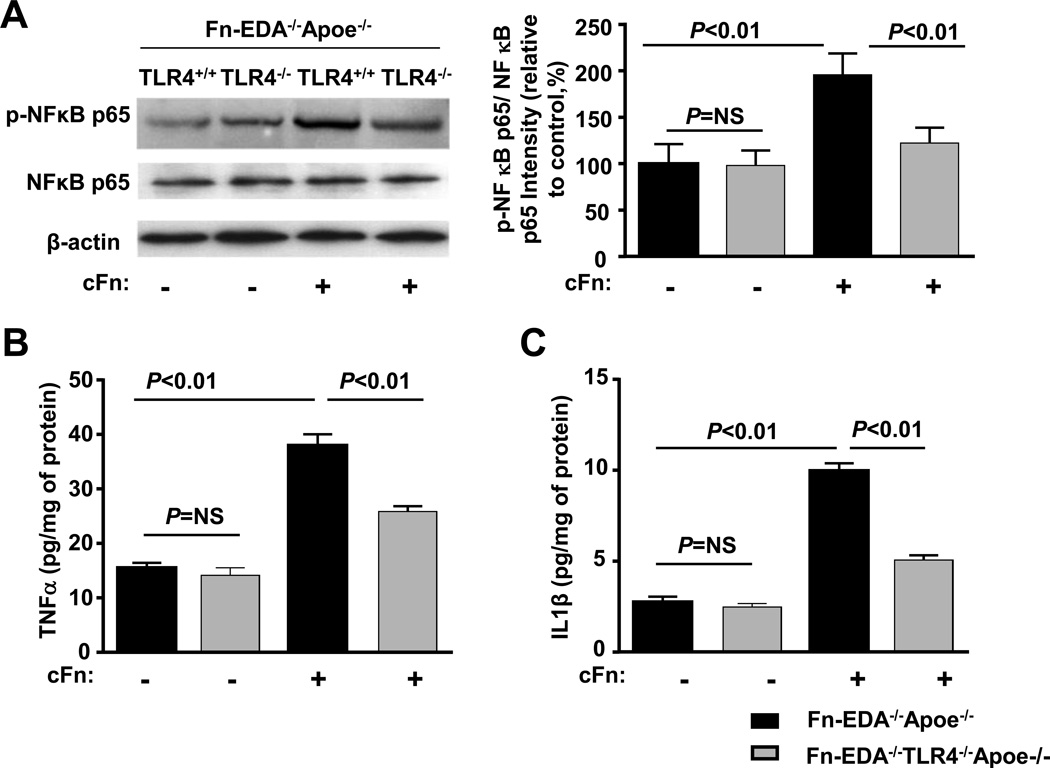
Figure 7.
Fn-EDA potentiates canonical NFκB signaling via TLR4. Bone marrow-derived neutrophils from Fn-EDA−/−Apoe−/− and Fn-EDA−/−TLR−/−Apoe−/− mice were stimulated with 20 ng phorbol myristate acetate in the presence or absence of cFn (10µg/well) for 24 hours. Left panel shows representative immunoblots of phospho- NFκB p65 and total NFκB p65. β actin was used as a loading control. The right panel represents quantification of intensity of phospho-NFκB p65 to total NFκB p65 in the presence or absence of cFn. Data are presented as mean ± SEM. N = 5/group. B &C. Quantification of TNFα and IL-1β by ELISA. Data are presented as mean ± SEM. N = 5/group.

