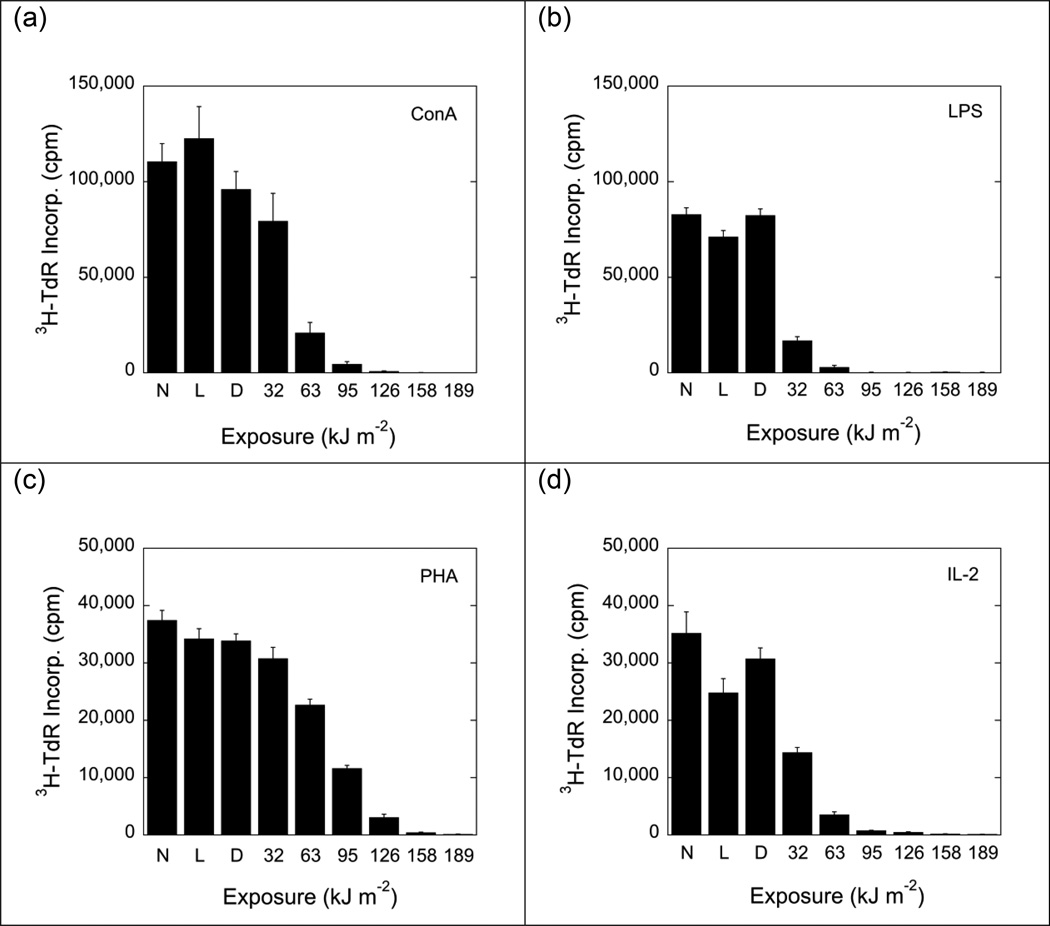
Fig. 2.
Proliferative responses (incorporation of 3H-thymidine) of non-treated and photoinactivated B6D2F1/J spleen cells to Concanavalin A (ConA; 2.5 µg ml−1), phytohemagglutinin (PHA; 15 µg ml−1), lipopolysaccharide (LPS; 5 µg ml−1), and interleukin-2 (IL-2; 25 U ml−1). Data represent means ± standard errors (SE) of 9 determinations derived from 3 independent experiments. Background activity (incorporation of 3H-thymidine by non-stimulated cells) was 3500 to 4500 cpm. N: Cells exposed to neither dye nor light. L: Cells exposed to light (189 kJ m−2) but no dye. D: Cells exposed to dye (15 µg ml−1) but no light. Suppressions of proliferative responses were statistically significant (p<0.05; 2-tailed Student's t-test) for cells exposed to MC540 and light fluences of ≥63 kJ m−2 (panels a and c) and for cells exposed to MC540 and light fluences of ≥32 kJ m−2) (panels b and d).

