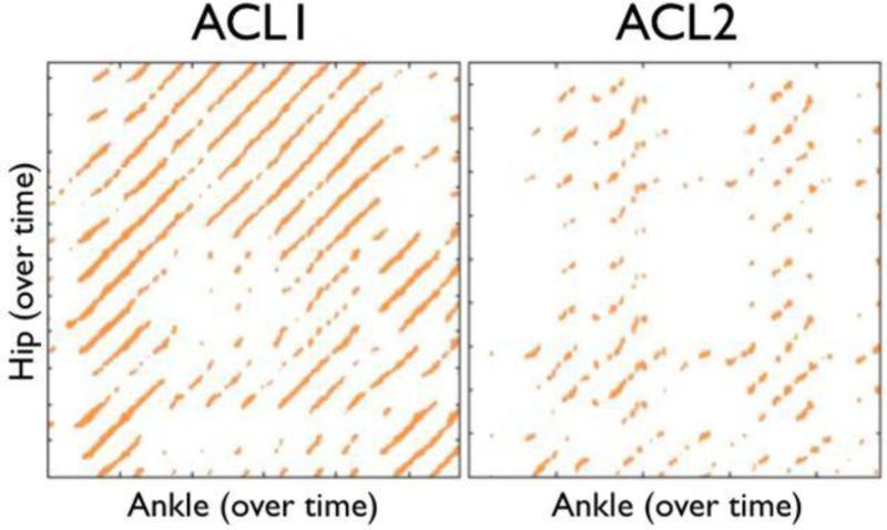
Figure 4.
Sample cross-recurrence plots for a single ACLR athlete who did not go on to second injury (ACL1—left) and an ACLR athlete who did go on to a second injury (ACL2—right). Note the degradation in the structure of the darkened pixels in the ACL2 plot compared to the ACL1 plot. The ACL2 athlete exhibits less tightly coupled coordination patterns and a qualitative breakdown in coordination.

