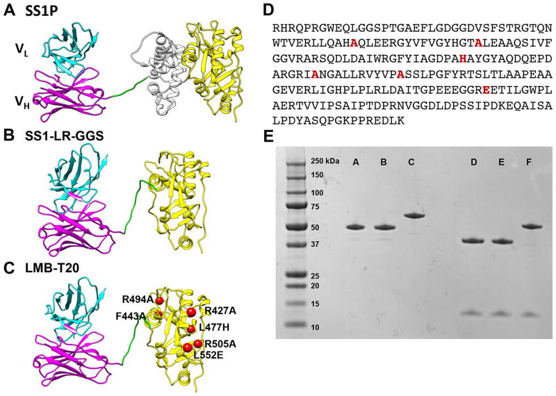
Figure 1.
Structural models of RITs. SS1P consists of a disulfide-stabilized heavy chain Fv polypeptide (VH) (cyan) and light chain Fv polypeptide (VL) (magenta) that are coupled to a 38-kDa fragment of PE38. (A) VH is recombinantly conjugated to the toxin fragment, which is composed of domain II (gray) and domain III (yellow). (B) SS1-LR-GGS. The SS1 Fv is conjugated to domain III with GGS linker between the furin cleavage site and domain III. (C) LMB-T20. Six point mutations were introduced into the scaffold of SS1-LR-GGS to diminish T cell epitopes. (D) Amino acid sequence of the toxin in LMB-T20. (E) SDS gel (4–20%) run under non-reducing conditions (lanes A, B and C) and reducing conditions (D, E and F) show the size and purity of the variant immunotoxins. Lanes A and D, LMB-T20, lanes B and E, LR-GGS and lanes C and F, SS1P.

