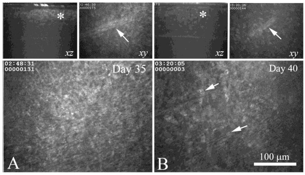
Figure 3.
In vivo confocal microscopy of the same eye from a CJLAT latently infected rabbit at Day 35 (A) and Day 40 (B). Upper left panels in A and B shows XZ projection through the same sub-clinical foci (asterisk), upper right panel shows the a nerve (arrow) that was located immediately below the sub-clinical foci in the same region. Lower panel shows the anterior stroma in the region of the sub-clinical foci, immediately below the basement membrane. Note that the sub-clinical foci appear less infiltrated (arrows) at Day 40 compared to Day 35 suggesting resolution of the lesion.

