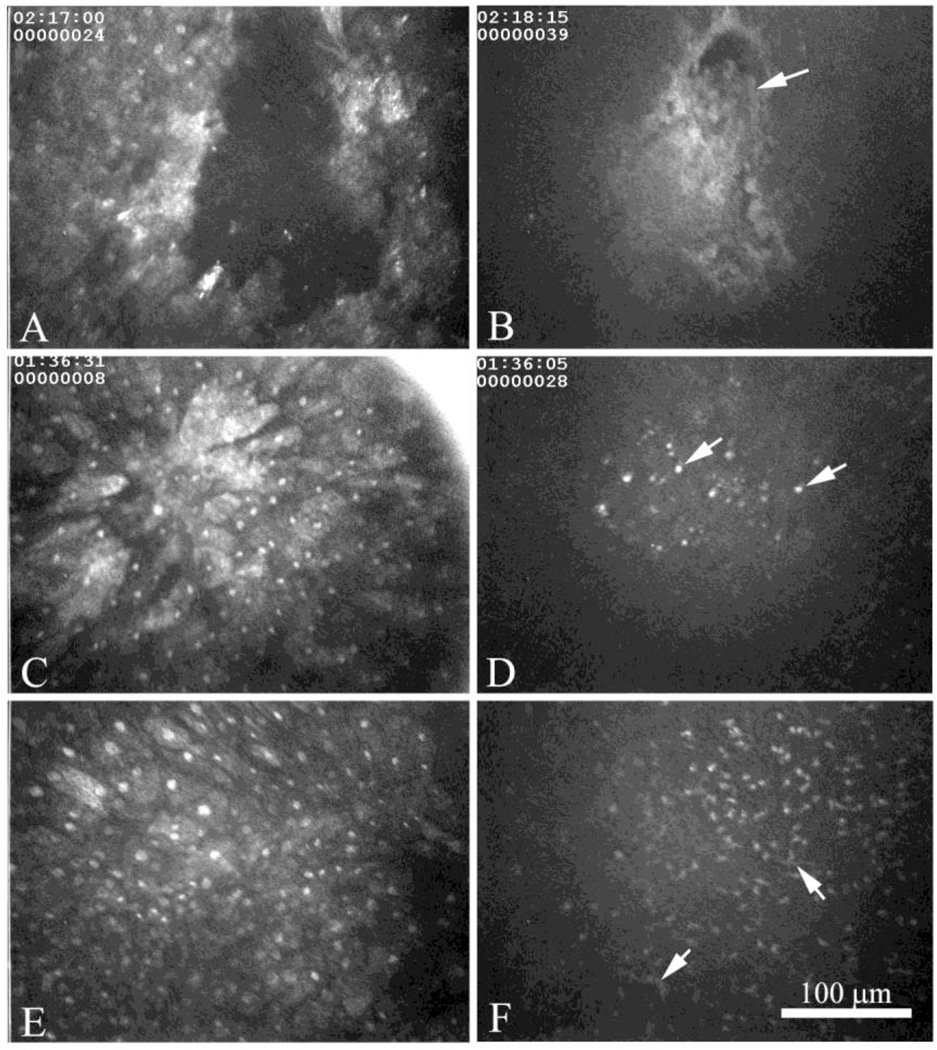
Figure 4.
In vivo confocal microscopic images of the surface epithelium (A, C, and E) and basement membrane (B, D, and F) showing epithelial erosion (A and B), migrating epithelium (C and D) and disorganized surface epithelium (E and F). At the level of the basement membrane epithelial erosions appear to contain necrotic basal epithelial cells (B, arrow), while regions of regenerating epithelium (C and E), were associated with varying degrees of small cell infiltrates (D and F, arrows).

