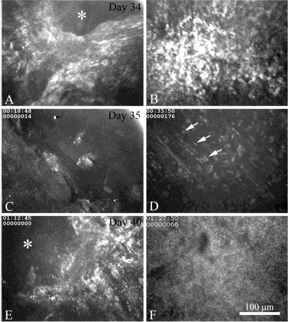
Figure 5.
In vivo confocal microscopic images of active recurrent HSK in the same eye beginning at Day 34 (A and B) and extending to Day 35 (C and D) and Day 40 (E and F). Images are taken at the level of the corneal surface (A, C, and E) and anterior corneal stroma (B, D, and F) in the same 3D data set. The first initiation of recurrent HSK at Day 35 was associated with the appearance of an epithelial lesions and exposure of the basement membrane (A, asterisk) that immediately overlaid a sub-clinical foci (B). Progression of recurrent HSK at Day 36 was associated with persistent epithelial erosion (C) and the infiltration of the anterior stroma with acute inflammatory cells (D, arrows). Four days later at Day 40, persistent epithelial erosion (E, asterisk) was associated with massive polymorphonuclear cell infiltration (F).

