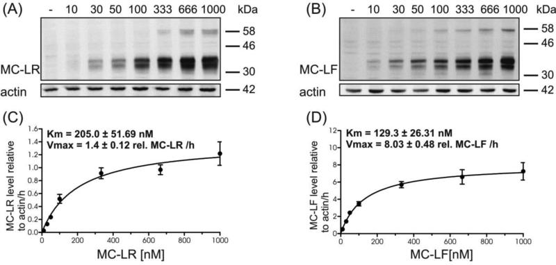
Fig. 4.
Concentration dependent uptake of MC-LR and MC-LF (10 nM-1000 nM) mediated by zfOatp1d1 using immunoblotting with anti-ADDA antibody. HEK293-zfOatp1d1 cells were exposed to increasing concentrations of MC-LR for 3 h (A) and MC-LF for 30 min (B), β-actin served as loading control. Quantification of transported MC-LR (C) and MC-LF (D) was carried out using anti-ADDA Westernblot and densitometric analysis. Vmax and Km were obtained by fitting the data from the greyscale analysis to the Michelis-Menten equation. Data are means ± SEM from three independent experiments.

