Figure 7.
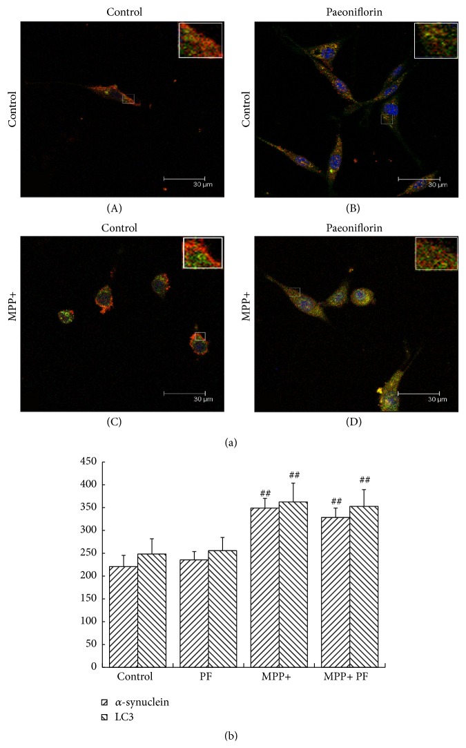
α-synuclein and LC3 colocalization in PC12 cells. Immunostain of α-synuclein (green) and LC3 (red) (a) and statistical analysis of optical density measurements (b), under normal conditions (A) and Paeoniflorin treatment (B), did not affect α-synuclein and LC3 and their colocalization; after MPP+ treatment (C), the signals of α-synuclein and LC3 were increased and lost their colocalization (more obvious in the enlarged figures). LC3 mainly aggregated in the peripheral cytoplasm but α-synuclein was distributed throughout the cell body. After MPP+ treatment and Paeoniflorin (D), the signals of α-synuclein and LC3 were both decreased and colocalization remained evident. Scale bar: 30 um. Quantification of immunostain results showed Paeoniflorin treatment did not affect fluorescence intensity of α-synuclein and LC3 in normal growth state (P > 0.05) but decreased the fluorescence intensity of α-synuclein and LC3 in MPP+ group. Values represent mean ± SEM (n = 5). # P < 0.05, ## P < 0.01 versus control group.

