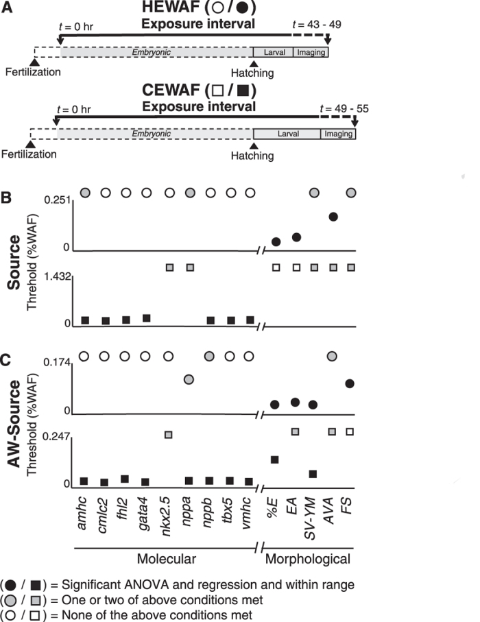
Figure 4. Comparison of threshold %WAF concentrations across molecular and morphometric cardiotoxicity indicators determined by linear regression for Source and AW-Source oils.

(A) Schematic depicting experimental design showing length of exposure, time of hatching, and period of imaging/tissue collection for HEWAF and CEWAF experiments. (B) Threshold %WAF values following exposure to Source 948 HEWAF (0–0.251%; top) and CEWAF (0–1.432%; bottom) preparations. (C) Threshold %WAF values following exposure to AW-Source oil HEWAF (0–0.174%; top) and CEWAF (0–0.247%; bottom) preparations. Molecular (amhc, cmlc2, fhl2, gata4, nkx2.5, nppa, nppb, tbx5, vmhc) and morphological (%E, EA, SV-YM, AVA, FS; see Methods) indicators are presented on left and right of X-axis break, respectively. Circles, HEWAF; squares, CEWAF; filled black symbols met all three of the following conditions: 1) significant one-way ANOVA (p < 0.05), 2) significant log-linear regression (p < 0.05), and 3) threshold %WAF within exposure range empirically tested (e.g., Supplemental Materials: Figure S5A); filled gray symbols met one or two of the above conditions; open symbols met none of the above conditions. Indicators with symbol above Y-axis maximum denotes non-significant log-linear regression (p > 0.05) or threshold %WAF value outside range empirically tested.
