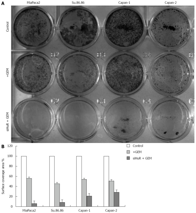
Figure 8.

Colony formation of pancreatic cancer cells (crystal violet). A: The colony formation (CV analysis) in 12-well plates; B: Quantitative CV analysis showed that cell surface coverage area was decreased in cells treated with GEM IC50 after HuR siRNA transfection compared with cells treated only with GEM IC50. HuR: Human antigen R; CV: Crystal violet.
