
Figure 1.
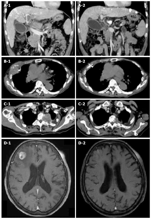
Tumor responses treated with Cyberknife. A1: CT scan of 59-year-old male with portal vein tumor thrombosis. The tumor is invading the portal vein from the main trunk to the 1st branch. The tumor diameter was 46 mm. A fiducial marker was implanted nearby; A2: Three months after irradiation with 35 Gy/5 fractions. The portal vein tumor thrombosis disappeared completely and the patient achieved CR; B1: CT scan of 85-year-old male with pleural HCC metastasis. The tumor diameter was 53 mm. A fiducial marker was implanted nearby; B2: Three months after irradiation with 30 Gy/5 fractions. The tumor disappeared completely and the patient achieved CR; C1: CT scan of 72-year-old male with thoracic spine HCC metastasis. Tumor is invading the left side of the thoracic spine at T2-3 causing bone destruction. The tumor diameter was 52 mm; C2: Three months after irradiation with 30 Gy/5 fractions. The tumor decreased to 33 mm (37% reduction in size) and the patient achieved PR; D1: T1-weighted MRI of 83-year-old female with brain HCC metastasis. There is a right frontal lobe lesion with gadolinium enhancement. The tumor diameter was 19 mm; D2: One month after irradiation of 20 Gy/1 fraction. The tumor disappeared completely and the patient achieved CR. CR: Complete response; CT: Computed tomography; HCC: Hepatocellular carcinoma.