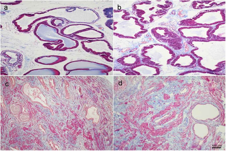
Figure 2. Histological examination of prostate.
Masson’s trichrome staining of prostate tissue. Prostatic SM cells were stained red, collagen fibers were stained blue and epithelial cells were stained orange. (a) Normal rat prostate. (b) BPH rat prostate. Hyperplastic prostate occurred mainly at the epithelial compartment and typical features of glandular hypertrophy was observed including increased acinus number, papillary fronds protruded into the glandular cavities and the epithelial layer thickened. (magnification × 100). (c) Normal human prostate. (d) Human BPH prostate. An obvious stromal hyperplasia was observed. (magnification × 100, n = 8 for each group).

