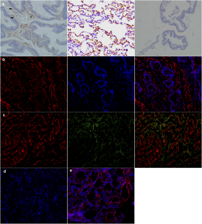
Figure 4. Immunolocalization of PDE5 and nNOS.
(a) Rat prostate. Left: PDE5 mainly distributed in fibromuscular stroma (black arrows) as well as in the endothelial and smooth muscle cells of blood vessels (black triangle). Middle: rat lung as positive control. Right: negative control. (magnification ×200). (b) Human prostate. Left: Cy3-immunofluorescence (red) indicates the PDE5 which was abundantly observed in the fibromuscular stroma. Middle: DAPI (blue) indicates nuclear staining. Right: Merged image. (magnification ×200). (c) Double immunofluorescence labeling of PDE5 and nNOS. Left: Cy3-immunofluorescence (red) indicates PDE5. Middle: Cy2-immuno fluorescence (green) indicates nNOS. Right: Merge image indicates the partly colocalization of PDE5 and nNOS in fibromuscular stroma. (magnification ×200). (d) Negative control by omitting the primary antibody. (magnification ×200). (e) Human lung tissue was used as positive control for PDE5. (magnification ×200, n = 8 from each group).

