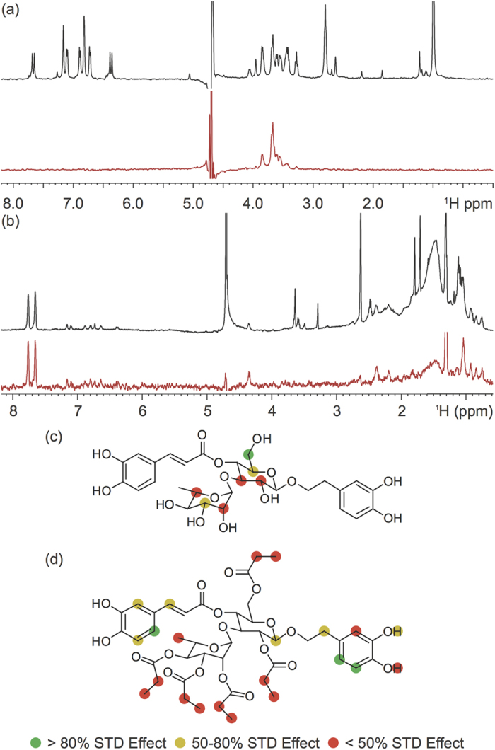
Figure 6. Ligand interaction with fibrillar Aβ42 by saturation transfer difference (STD) NMR (600 MHz).

STD NMR spectra of (a) Verbascoside and (b) VPP at a 250:1 ligand to peptide ratio using pre-assembled Aβ42 fibrils (1 μM). Comparison of the STD signal intensity (red) to the STD reference intensity (black) reflects the relative proximity of the corresponding proton to the Aβ42 fibril. The STD effect calculated from relative intensities was correlated to the structure of Verbascoside (c) and VPP (d) to generate group epitope maps.
