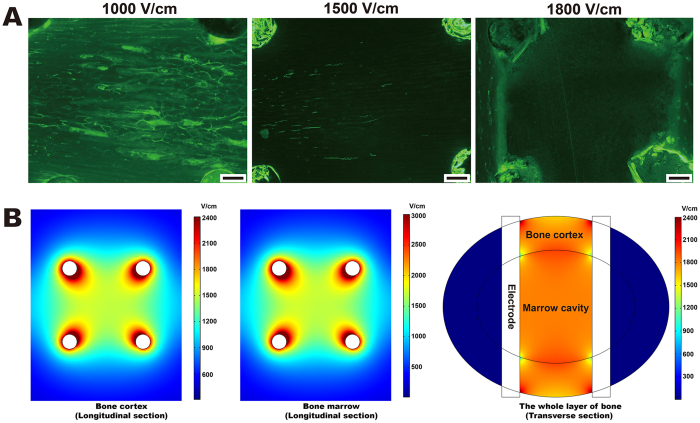
Figure 1. Determination of the effective ablation threshold.
(A) Effectiveness of IRE ablation using three different electrical intensities. One hundred twenty direct-current pulses at 1,000 V/cm, 1,500 V/cm or 1,800 V/cm were applied to animals in the threshold group. Calcein green labeling was observed 1 week after IRE ablation at 1,000 V/cm and at 1,500 V/cm, but no labeling was observed at 1,800 V/cm. Bars represent 500 μm. (B) Distribution of the electric field intensity at 1,800 V/cm. Pulses of 1,800 V/cm were output 6 times, including on four sides and two diagonals of the square configuration. The electric field distribution in a square configuration was non-uniform, as evidenced by the graduated color. In the longitudinal section, the lowest intensity occurred at the center of the square (1,610 V/cm in the cortex and 1,730 V/cm in the marrow). In the transverse section, the lowest intensity occurred at the intersection of the cortex, marrow and electrode (1,310 V/cm).

