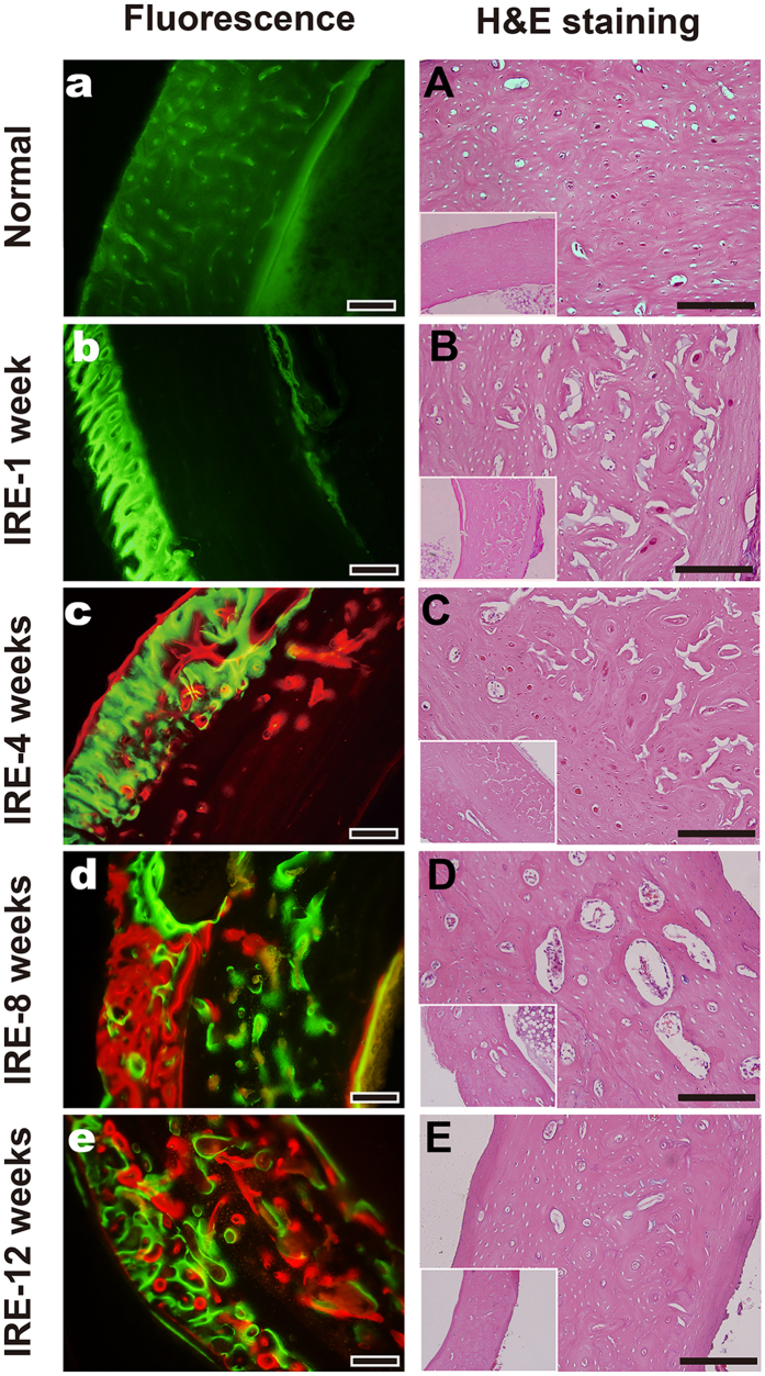
Figure 3. Histopathological assessment of the IRE-ablated bone with fluorescence and H&E staining.
(a–e): Fluorescent labeling of the target bone segment with calcein green or alizarin red in the corresponding period. (A–E): H&E staining of the target bone segment in the corresponding time period. Bars represent 200 μm.

