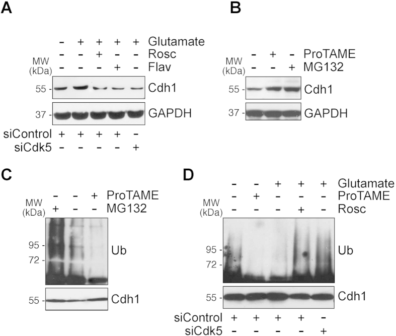
Figure 2. Cdk5 phosphorylates Cdh1 and triggers APC/C inhibition causing Cdh1 protein stabilization.
(A) Rat cortical neurons on day 4 in vitro were transfected with a siRNA against luciferase (siControl; 100 nM) or with siRNA against Cdk5 (siCdk5; 100 nM) for 3 days. Neurons were then treated with glutamate (100 μM, 5 min) and were further incubated in culture medium, supplemented with Cdk inhibitors, 10 μM roscovitine (Rosc) and 1 μM flavopiridol (Flav), APC/C inhibitor, ProTAME (10 μM), and proteasome inhibitor, MG132 (10 μM), for 4 h. Cdh1 protein levels were analyzed by Western blotting. GAPDH protein levels were used as loading control (B) Neuronal were treated with ProTAME (10 μM), and MG132 (10 μM), for 4 h, and Cdh1 protein levels were analyzed by Western blotting. (C) Neuronal extracts were immunoprecipitated with anti-Cdh1 antibody and analyzed by Western blot for ubiquitin (Ub). (D) Rat cortical neurons were treated as in (A) and neuronal extracts were immunoprecipitated with anti-Cdh1 antibody and analyzed by Western blot for ubiquitin (Ub). In all cases a representative western blot is shown out of three. The relative protein abundance is shown in Supplementary Fig. 1.

