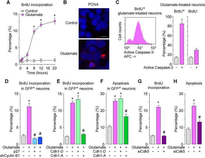
Figure 4. Cdk5-mediated Cdh1 phosphorylation induces cell cycle entry leading to neuronal apoptosis in excitotoxicity.
Rat cortical neurons were treated with glutamate (100 μM, 5 min) and were further incubated in culture medium for 4–20 h. (A) Bromo-d-Uridine (BrdU) incorporation was measured by flow cytometry. (B) At 20 h after glutamate treatment, PCNA (proliferating cell nuclear antigen) was detected by immunocytochemistry. Microphotographs reveal that glutamate induced nuclear PCNA expression in neurons. Scale bar = 10 μM. (C) Immediately after glutamate stimulation, neurons were incubated in culture medium containing 10 μg/mL BrdU, for 20 hours. Neurons were then immunostained with BrdU and active caspase-3, which were detected by flow cytometry. Most (85%) of the BrdU+ neurons cells expressed active caspase-3. (D) Neurons were transfected with pSuper-neo.gfp-Cyclin B1 (expressing both shCyclin B1 and the enhanced GFP) or pIRES2-EGFP mammalian expression vector co-expressing p27 and the enhanced GFP. Control cells were transfected with pSuper-neo.gfp (shControl). Neurons were then treated with glutamate (100 μM, 5 min) and were further incubated in culture medium for 20 h. Flow cytometry analysis for BrdU incorporation was performed in transfected (identified by GFP fluorescence) neurons. (E,F) Neurons were transfected with pIRES2-EGFP mammalian expression vector co-expressing the enhanced GFP and either the phosphomimetic (Cdh1-P) or phosphodefective (Cdh1-A) forms of Cdh1. Control cells were transfected with empty vector (pIRES2-EGFP). Neurons were then treated with glutamate (100 μM, 5 min) and were further incubated in culture medium for 20 h. BrdU incorporation and neuronal apoptosis were measured in GFP+ (transfected) neurons by flow cytometry. (G, H) Neurons were transfected with siRNA against luciferase (100 nM; siControl) or with siRNA against Cdk5 (100 nM; siCdk5) for 3 days. Neurons were then treated with glutamate (100 μM, 5 min) and were further incubated in culture medium, for 20 hours. BrdU incorporation and neuronal apoptosis were measured by flow cytometry. In all cases, the represented values are means ± S.E.M. (n = 3 independent neuronal cultures). *p < 0.05 versus untreated (Control) neurons. #p < 0.05 versus Glutamate-treated neurons.

