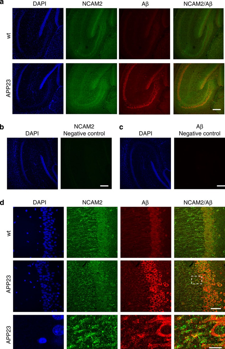
Figure 7. NCAM2 co-localizes with Aβ1-42 in brains of APP23 transgenic mice.
Low-magnification confocal images of the hippocampus (a) and high-magnification images of the CA1 region of the hippocampus (d) of 9-month-old wild-type (wt) and APP23 mice (APP23) are shown. Brain sections were immunolabelled with antibodies to NCAM2 and Aβ and counter stained with DAPI. Lower panel in d shows a blow-up of the area outlined with dashed lines. Note accumulations of Aβ co-localizing with clusters of NCAM2 along dendrites of neurons. Negative controls (labelling performed without primary antibodies) are shown in b,c. Scale bar=200 μm (a–c), 40 μm (d), 10 μm (d, blow-up image).

