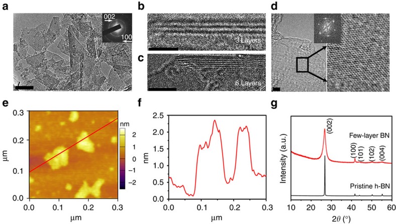
Figure 2. Exfoliated few-layer BN.
(a) TEM image of few-layer BN, with the selected-area electron diffraction pattern (inset) indicating a layered BN structure. Scale bar, 50 nm. (b,c) HRTEM images of the edge folding of two few-layer BN sheets with three and six BN layers, respectively. Scale bars, 2 nm (b) and 5 nm (c). (d) HRTEM and the fast Fourier transform images of a few-layer BN sheet. Scale bar, 2 nm. (e,f) Atomic force microscopy image and corresponding line-scan profile of few-layer BN. (g) XRD patterns of few-layer BN and pristine h-BN.

