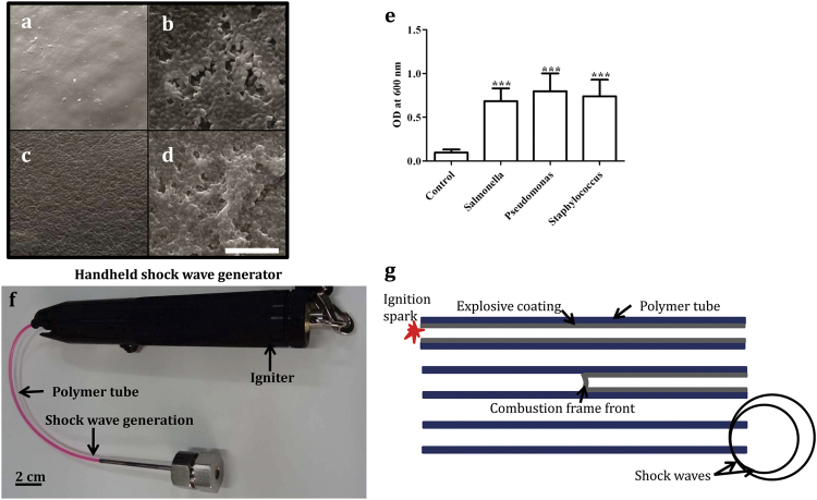
Figure 1. Biofilm formation on urinary catheters.
(a–d) Scanning electron micrographs (SEM) of bacterial biofilms on sections of urinary catheter. The micrographs were taken at the same magnification and the scale bar in (d) is 10 μm. (a) Untreated catheter surface. (b) Catheter surface with S. Typhimurium biofilm. (c) P. aeruginosa biofilm. (d) S. aureus biofilm. (e) Crystal violet staining of biofilms caused by the different bacteria on equal amounts of catheter substrate. Statistical significance was calculated using One-way ANOVA. Asterisks indicate statistical significance as follows: (***p < 0.001). Error bar–mean ± SD. (f) Photograph of hand held shock wave generator. (g) Schematic of hand held shock wave generator. The igniter is used for the ignition of the polymer tube by a spark generated by electrodes, explosive coating undergoing combustion and the combustion flame front travelling at 2000 m/s, and shock waves emanating from the open end of the polymer tube.

