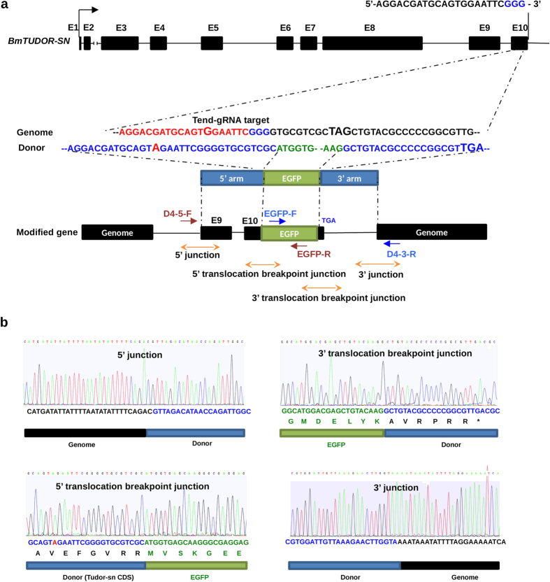
Figure 6. Sequencing of the modified BmTUDOR-SN gene.
(a) A schematic representation of the BmTUDOR-SN gene and donor consisting of homologous arms and EGFP coding region. The gRNA targeting sequence was marked by red, and PAM was marked by blue. A guanine for modification was bold in the gRNA-targeting site in the genome sequence. The mutated nucleotide adenine was bold and red-marked in the donor sequence. The stop codons in genome or donor were bold. DNA fragments for sequencing were amplified by the primer sets, red-marked primers for 5′ junction and 5′ translocation breakpoint junction, blue-marked primers for 3′ junction and 3′ translocation breakpoint junction. (b) Chromatograms of DNA sequences of 5′ junction, 5′ translocation breakpoint junction, 3′ junction and 3′ translocation breakpoint junction in the modified BmTUDOR-SN gene. The homologous arms were marked by blue. EGFP coding sequence was marked by green. Genome was marked by black.

