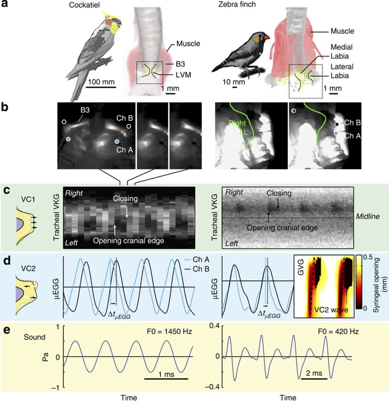
Figure 3. MEAD theory explains syringeal sound production in neognaths.
(a) Three-dimensional syrinx geometry of the cockatiel (Nymphicus hollandicus, ventral view) and zebra finch (Taeniopygia guttata, dorsal view) showing bone (grey), LVM (yellow) and muscles (pink). Muscle is rendered transparent in zebra finch right hemisyrinx to show vibratory tissue. (b) High-speed imaging stills of frontal transilluminated frontal views of the syrinx at two different phases during the oscillatory cycle at view and location indicated with a dotted box in a. While transillumination does not provide detail on motion in the cockatiel syrinx, it provides sufficient detail to identify inner edges of vibratory tissue in female zebra finches (green). Circles indicate location of μEGG electrodes (cyan, channel A; black, channel B). (c) Tracheal endoscopic VKG shows opening and closing events and thus evidence for medio-lateral vibrations (VC1). In the zebra finch sound production was induced in the right hemisyrinx. (d) Evidence for caudo-cranial component (VC2) of the tissue wave as demonstrated by μEGG phase delays for cockatiel and zebra finch and glottovibrogram analysis for zebra finch. (e) Synchronised acoustic waveforms.

