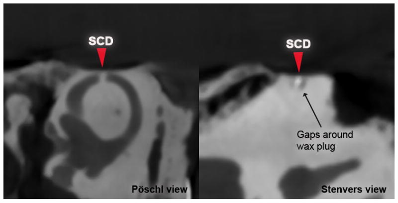
Figure 4. Partial occlusion of SSC.

Pöschl (left) and Stenvers CT (right) views of a specimen after repair of 0.5 mm dehiscence using radiopaque wax. Arrows mark location of SCD.

Pöschl (left) and Stenvers CT (right) views of a specimen after repair of 0.5 mm dehiscence using radiopaque wax. Arrows mark location of SCD.