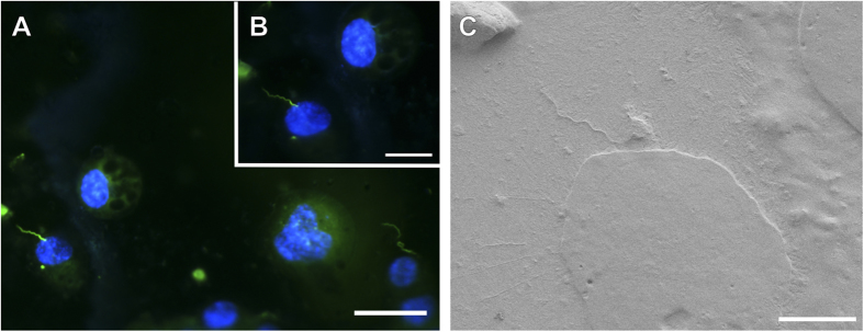
Figure 3. Demonstration of fluorescence preservation after deposition of Pt/Pd layer on the sample followed by SEM examination under cryo-conditions.
Association of B. burgdorferi-GFP (green) with mouse neuroblastoma cells. Cells were counterstained with Hoechst 33342 (blue). Images acquired before (A) and after (B) cryo-SEM examination. (B) Image taken after 20 minutes ice sublimation and deposition of Pt/Pd layer on the sample surface. (C) Cryo-SEM micrograph of the spirochete-cell interaction shown in (A) and (B). Scale bars: (A) 50 μm, (B) 25 μm and (C) 10 μm.

