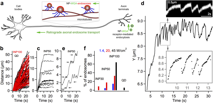
Figure 1. Nanoparticle assisted optical tethering of endosomes in axons.
(a) Schematic of the retrograde axonal transport of nanoparticle-WGA endosomes from axon terminals to the cell bodies in neurons. (b) Unperturbed retrograde transport trajectories of QD-endosomes (black, 45 W/cm2, 32fps) and INP100-endosomes (red/gray, 1.4 W/cm2, 10fps). (c) Retrograde INP50-endosomes (19 W/cm2, 32fps) stochastically becoming stationary within the imaging field of view. (d) Gradual stalling and fast reversals (jumps) exhibited by an affected INP-endosome. Inset zooms in on a few jumps with the corresponding kymograph is also shown. (e) Laser-affected INP50-endosomes exhibiting jumps at different locations along the axon. (f) Percentage of laser-affected endosomes, for different nanoparticles at varying laser powers.

