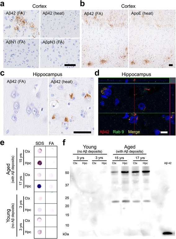
Fig. 1.

Aβ deposits in cat brains. a Aβ42 aggregates are detected in the parenchyma of the cerebral cortex with anti-Aβ42 antibody (12 F4) on formic acid (FA)-pretreated sections but not on heat-pretreated sections. These aggregates are not detected with antibodies against the N-terminus of human Aβ (AβN1 and AβpN3). b Aβ42 aggregates in the cerebral cortex colocalized with ApoE. c Heat pretreatment revealed intracellular Aβ42 aggregates in the pyramidal cells of the hippocampus but not in the cortex. d Some of the intracellular Aβ42 (red) aggregates colocalized with Rab9 (green). Black bars = 50 μm, white bar = 10 μm. e Dot blot analysis of SDS fractions and FA fractions of cortex (Ctx) and hippocampus (Hpc) of young cats and aged cats. Aβ oligomers were detected with A11 antibody, predominantly in the SDS fraction from the hippocampus of aged cats. f Western blotting analysis of the SDS fraction of the Ctx and Hpc of young cats and aged cats. Two distinct bands were detected with anti-Aβ antibody 6E10 in the brains of aged cats: approximately 24 kDa and 54 kDa, indicating Aβ hexamers and dodecamers, respectively
