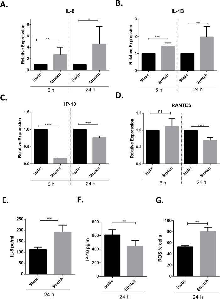
Figure 3. Cyclic stretch activates a pro-inflammatory response and enhances oxidative stress.
(A–D) VA10 cells were subjected to stretch for 6 and 24 h. The mRNA expression of genes encoding pro inflammatory cytokines IL-8 (A), IL-1β (B) and chemokines IP-10 (C), RANTES (D) was measured with q-RT PCR (n = 3, mean ± S.E.). Relative expression levels (y-axis) in static cells were defined with an arbitrary value of ‘1’ and changes relative to this value in stretched samples are represented. (E–F) The protein expression of IL-8 (E) and IP-10 (F) from cultured supernatants was measured with ELISA. VA10 cells were stretched for 24 h and ELISAs were performed (n = 3, mean ± S.E.). (G) Oxidative stress was measured with CellROX green reagent. VA10 cells were subjected to stretch for 24 h. CellROX dye (5 µM) was added 30 min before the end of stretching. The cells were then harvested and analyzed by flow cytometry. The data is represented as percentage positive CellROX (ROS) cells before and after cyclic stretch (n = 3, mean ± S.E.) (ns indicates non-significant; p < 0.05, ∗; p < 0.01, ∗∗; p < 0.001, ∗∗∗; p < 0.0001, ∗∗∗∗).

