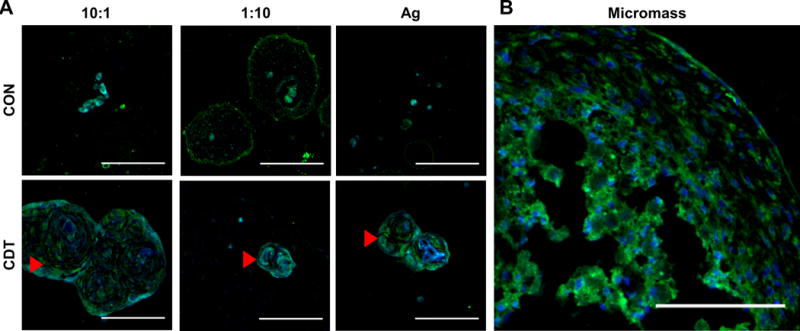
Figure 9.

Collagen type II labeling of PEC-Ag microbeads cultured in control (CON) or chondrogenic (CDT) medium. Green = Collagen type II (FITC secondary antibody). Blue = DAPI stain. Arrows show masses of ECM containing Collagen type II. As with the Alcian Blue-PAS histology (see Fig.8), large agglomerations of extracellular matrix were found in the 10:1 microbeads. Scale bar = 100 μm.
