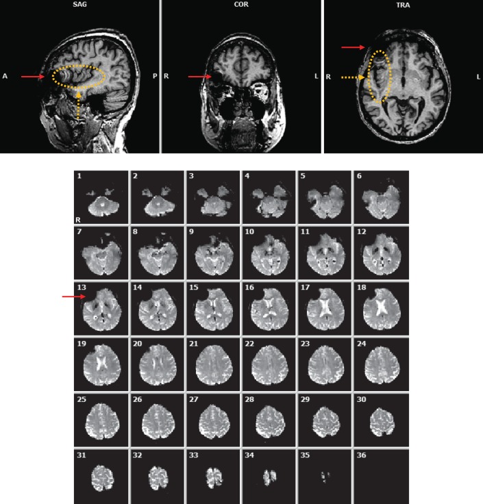
Figure 1.
Top: Anatomical (MP-RAGE) image of subject A2 displaying local and nonlocal forms of artifact. Bottom: 36 functional (EPI) image slices showing presence of an artifact near the right eye (left side of images). Red solid arrows: local artifact resulting from the presence of the device implanted in the subject's right eye. Orange dashed arrows and ellipse: nonlocal artifact in the right hemisphere extended along the phase encoding direction (AP).

