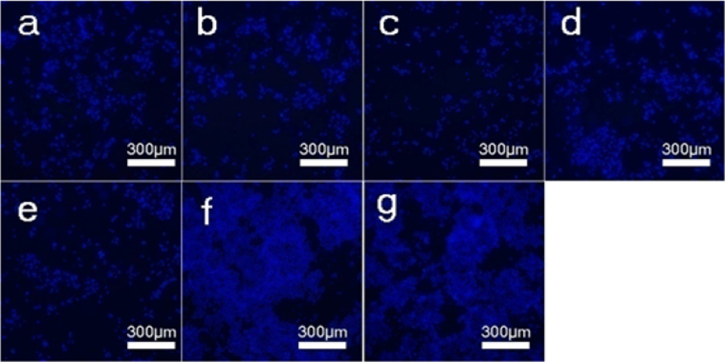
Figure 7.
Confocal fluorescence images of MDA-MB-231 breast cancer cells after incubation with different DOX-loaded microspheres at a DOX concentration of 10 µg/mL for 5 days. The samples were as follows: (a) DOX-loaded M-0, (b) DOX-loaded M-300, (c) DOX-loaded M-305, (d) DOX-loaded M-310, (e) Free DOX, (f) DOX-free vaterite microspheres (M-0), and (g) blank plate as control. The blue (DAPI) is living cancer cells.

