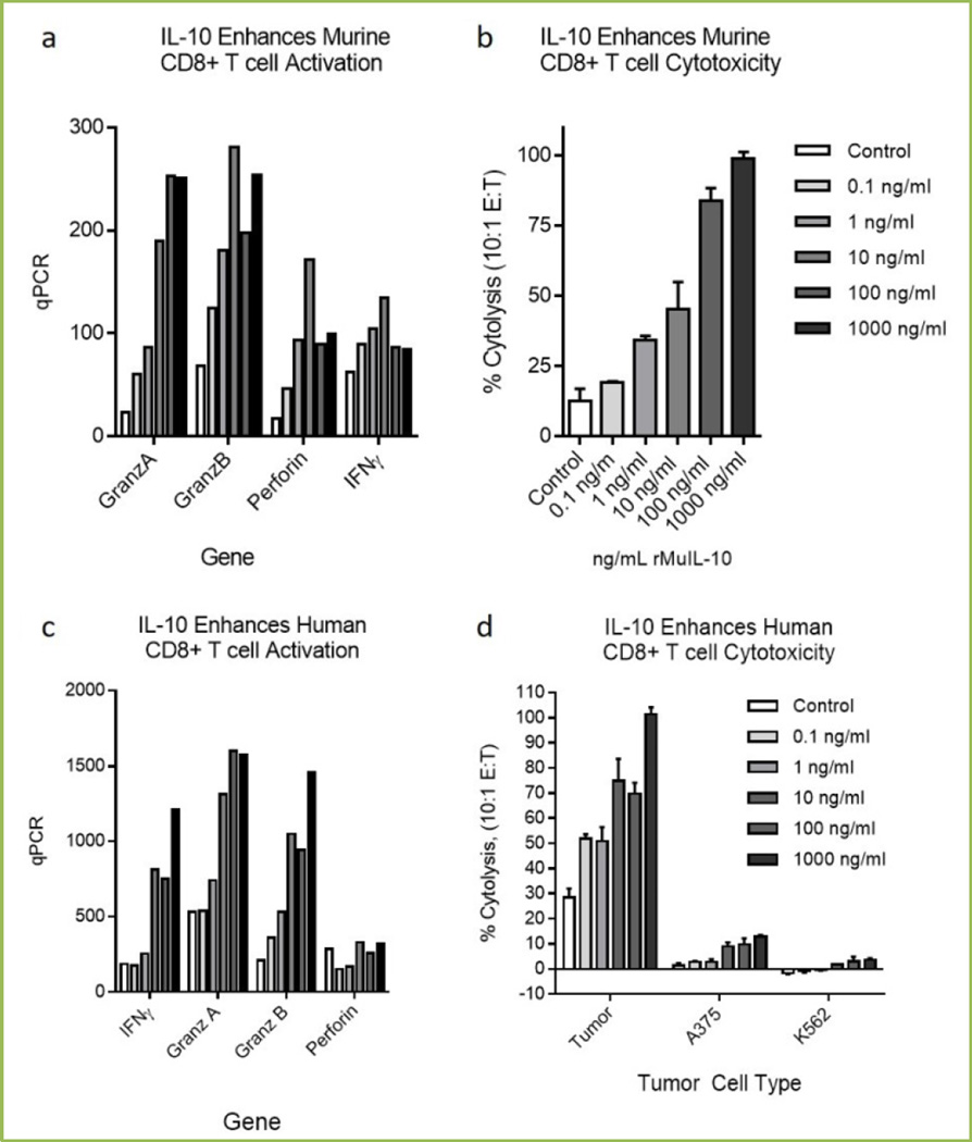
Fig 1. IL-10 exposure directly stimulates murine and human CD8+ T cell cytotoxic function.
Murine OT1 T cells isolated by magnetic (Miltenyi) bead isolation were stimulated and exposed to species specific IL-10 for 3–5 days. Cells were assessed for cytotoxic mRNA regulation (1a) and for cytolytic function (1b). OT1 cells were exposed at 10:1 effector to target ratio and cytolysis determined by CytoTox96 (Promega) LDH release. Target cells were PDV6 squamous tumor cells pulsed with SIINFEKL for 24 hours. Cytolysis was assessed after 4 hours. Human CD8+ T cells were isolated by magnetic (Miltenyi) bead isolation (1c), stimulated and exposed to IL-10 for 3–5 days. Cells were assessed for cytotoxic mRNA regulation (1c) and for cytolytic function (1d). Tumor antigen specific CD8+ T cells were isolated from surgically resected human melanoma tumors and stimulated with irradiated autologous tumor cells in the presence of 100U/mL IL-2 (Centocor) and rested for 5 days with IL-10. Activated CD8+ T cells were exposed at 10:1 effector to target ratio and cytolysis of Cr51 labeled autologous tumor cells, A375 (ATCC) or K562 (ATCC) assessed after 4 hours.

