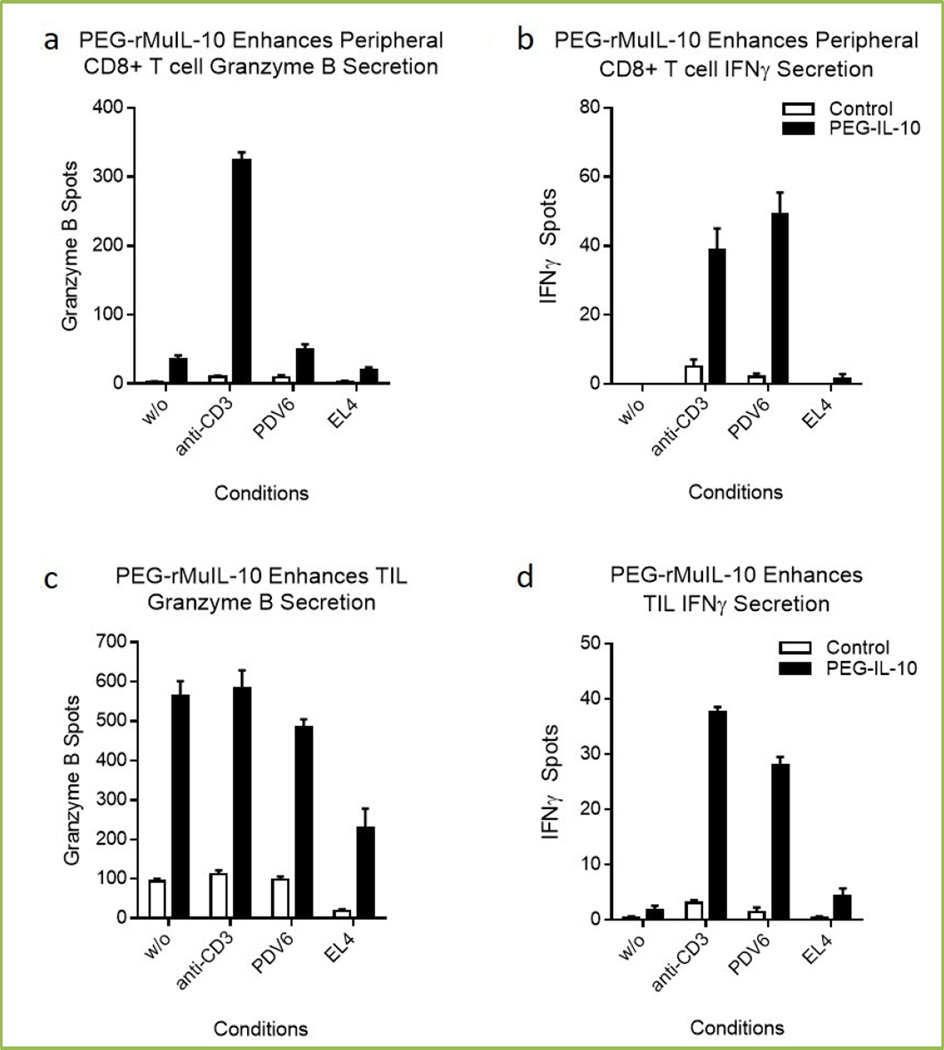
Fig 2. Effect of PEG-rMuIL-10 dosing on peripheral and intratumoral CD8+ T cell cytotoxicity.
These ELISPOTs (R&D Systems) were generated by magnetic (Miltenyi) bead isolation of 1000 CD8+ T cells from PBMC or mechanically disrupted and enzyme digested PDV6 (Schering Plough) squamous tumors. CD8+ T cells were exposed for 24 hrs without any secondary stimulus, (w/o), or 1 µg/mL soluble anti-CD3 (eBiosciences), 100 PDV6 squamous tumor or EL4 (ATCC) (as negative control) tumor cells. Spots were quantified with ImmunoSpot Software.

