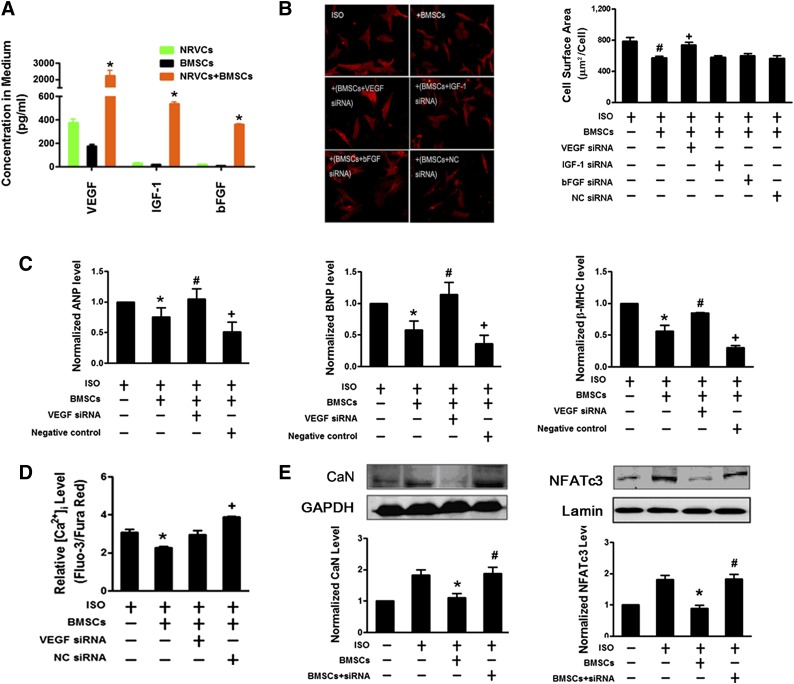
Figure 3.
VEGF involves the reversal of cardiomyocyte hypertrophy by BMSCs. (A): Coculture of neonatal rat ventricular cardiomyocytes (NRVCs) with BMSCs increases the contents of VEGF, bFGF, and IGF-1 in the medium. ∗, p < .05 versus NRVC monoculture or BMSC monoculture. (B): Silencing of VEGF, but not bFGF and IGF-1, by siRNAs (300 pM) in BMSCs abolishes the amelioration of ISO-induced enlargement of cardiac cell size by BMSCs. BMSCs were pretreated with siRNAs by transfection and then cocultured with NRVCs. Left: Representative images of NRVCs (×200 magnification, at least 10 randomly selected fields in three separate experiments) with α-actinin staining. Right: Averaged cell surface area of NRVCs. The scrambled NC siRNA failed to produce the effects against BMSCs. siRNAs were transfected into BMSCs. (C): VEGF siRNA abolishes the inhibitory effects of BMSCs on the ISO-induced expression of hypertrophic marker genes ANP, BNP, and β-MHC mRNAs in NRVCs. (D): VEGF siRNA abolishes the inhibitory effects of BMSCs on the ISO-induced increase of [Ca2+]i in NRVCs. (E): VEGF siRNA abolishes the inhibitory effects of BMSCs on the ISO-induced upregulation and activation of CaN and nuclear NFATc3 proteins in NRVCs. The data were obtained from four independent experiments. ∗, p < .05 versus ISO; #, p < .05 versus BMSCs; +, p < .05 versus BMSCs transfected with VEGF siRNA. Abbreviations: ANP, atrial natriuretic peptide; bFGF, basic fibroblast growth factor; BMSC, bone marrow-derived mesenchymal stem cell; BNP, brain natriuretic peptide; CaN, calcineurin; Ctl, control; GAPDH, glyceraldehyde-3-phosphate dehydrogenase; IGF-1, insulin-like growth factor 1; ISO, isoproterenol; β-MHC, β-myosin heavy chain; NC, negative control; NFATc3, nuclear factor of activated T cells cytoplasmic 3; siRNA, small interfering RNA; VEGF, vascular endothelial growth factor.

