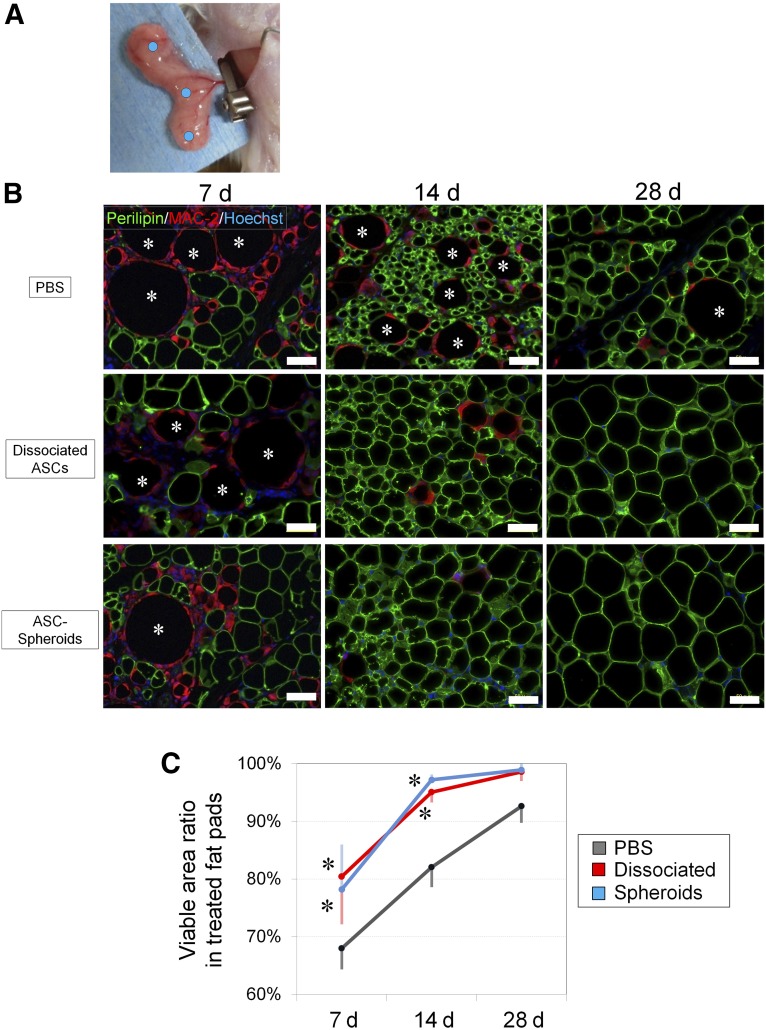
Figure 5.
Histological evaluation of cell-based treatments for ischemia-reperfusion injury of inguinal fat pad in SCID mice. (A): The main nutrient vessels arising from the femoral vessels were clamped with a microclip for 4 hours and then allowed to perfuse. Mice models were divided into three groups, and each group was treated with an injection of PBS, monolayer-cultured ASCs, or ASC spheroids at three points (blue dots) of the fat pad. (B): Immunohistochemistry for perilipin (viable adipocytes) and MAC-2 (macrophages) revealed that both monolayer-cultured ASCs and ASC spheroids improved the adipose tissue repair and regeneration after ischemia-reperfusion injury. Many dead adipocytes (perilipin-negative) surrounded by infiltrated M1 macrophages were detected at 7 days, indicating substantial tissue damage by the injury. At 14 days, adipose regeneration (perilipin-positive small adipocytes) was seen, and the tissue repair was almost completed by 28 days in samples treated with monolayer-cultured or spheroid ASCs. Asterisks show dead adipocytes. Scale bars = 50 μm. (C): At 7 and 14 days, samples treated with monolayer-cultured or spheroid ASCs showed an increased viable area ratio compared with PBS-treated samples, suggesting that the injury repair process was accelerated by the cell treatments. ∗, p < .05 compared with control (PBS). Abbreviations: ASC, adipose-derived stem/stromal cell; d, days; PBS, phosphate-buffered saline.

