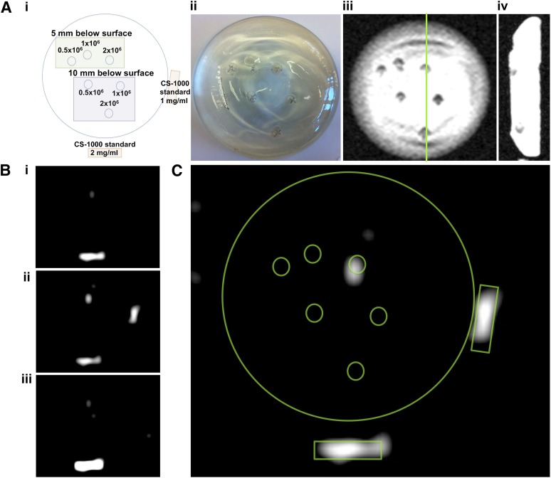
Figure 5.
19F 3-Tesla magnetic resonance imaging (MRI) of labeled stromal vascular fraction (SVF) cells in a silicone breast phantom. (Ai): Diagram outlines the six cell injection variables (0.5, 1.0, and 2.0 × 10⁶ cells injected at either 5- or 10-mm depth) and CS-1000 standards. (Aii): An optical image of the final injected breast implant phantom. Coronal (Aiii) and sagittal (Aiv) 1H MR images of the phantom show nonspecific contrast from injection sites. (Bi–Biii): Sequential 19F MR images of the phantom show MRI signal only for 2.0 × 10⁶ cells injected at a 5-mm depth. (C): This was confirmed by overlaying the cell and standard diagram on the slice from (Bii).

