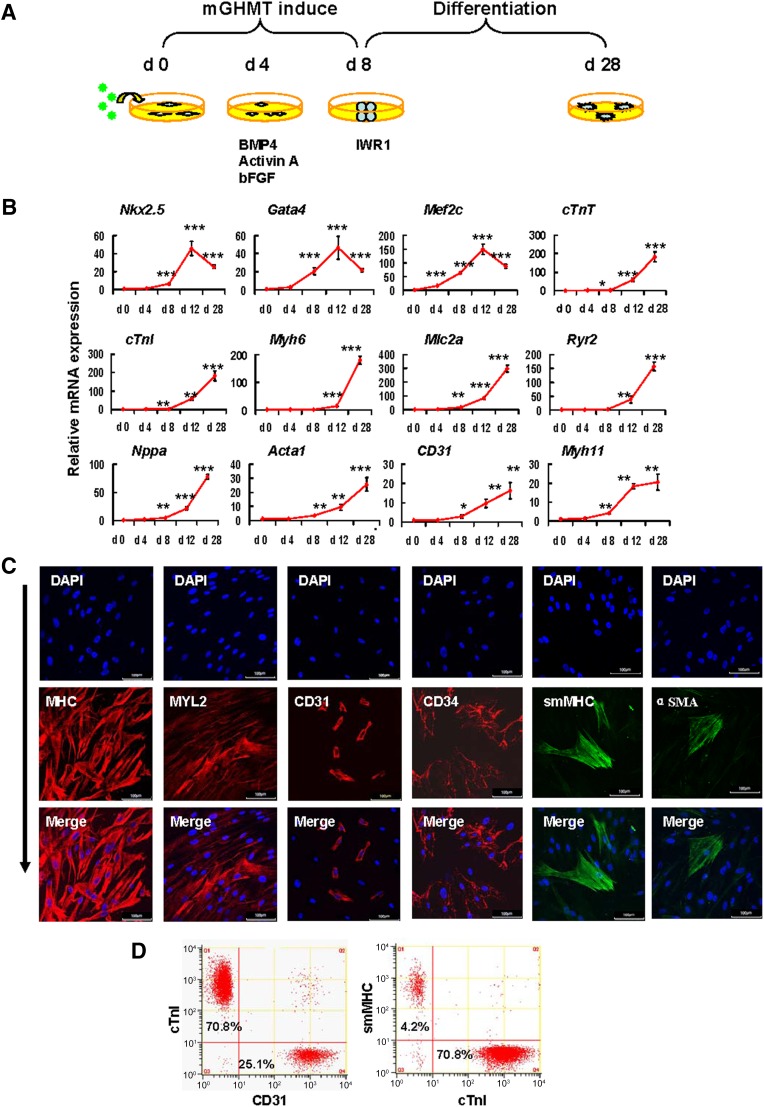
Figure 3.
Protein-induced cardiac progenitor cells (piCPCs) differentiated into three cardiac lineages: cardiomyocytes, endothelial cells, and smooth muscle cells. (A): Schematic representation of the strategy to differentiate piCPCs in differentiation medium with IWR1 factor. (B): Quantitative data of mRNA expression of cardiac lineage marker genes (∗, p < .05; ∗∗, p < .01; and ∗∗∗, p < .001 vs. day 0 control; error bars indicate SD; n = 3). (C): Immunofluorescent staining for MHC, MYL2, CD31, CD34, smMHC, and αSMA. The combination of the four factors, GHMT, induces abundant MHC and Myl2, and some expression of CD31 and smMHC 28 days after transduction. Nuclei were counter stained with DAPI. Scale bars = 100 μm. (D): Flow cytometry analysis for cTnI, CD31, and smMHC. mGHMT plus IWR1 significantly enhances cTnI expression, and, to a lesser extent, CD31 and smMHC expression. Abbreviations: αSMA, α-smooth muscle actin; BMP4, bone morphogenetic protein 4; cTnI, cardiac troponin I; cTnT, cardiac troponin T; d, day; DAPI, 4′,6-diamidino-2-phenylindole; GHMT, Gata4/Hand2/Mef2c/Tbx5; mGHMT, modified GHMT; MHC, myosin heavy chain; MYL2, myosin light chain 2; smMHC, smooth muscle myosin heavy chain.

