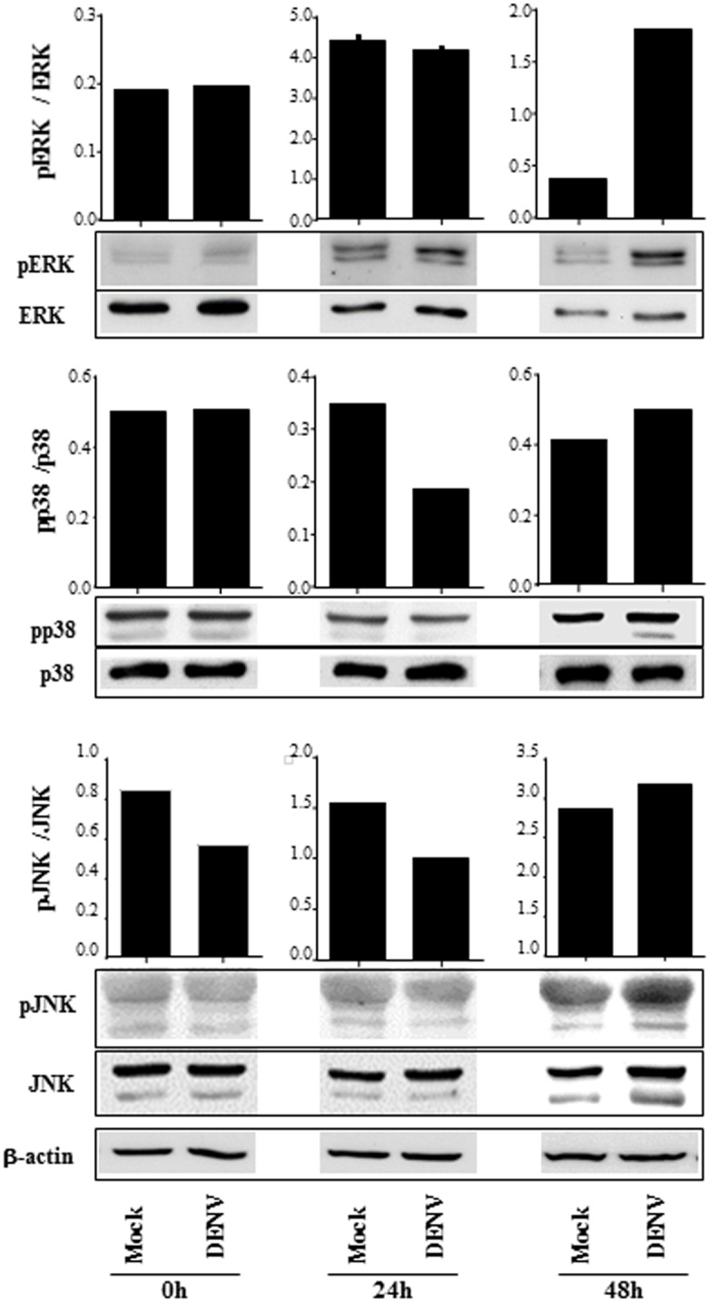
Fig 3. B cell infection by DENV promotes MAPK phosphorylation.

B lymphocytes were mock-treated or cultured with DENV2 (MOI = 1). The cells were harvested after 2h, 24h or 48h p.i., and the expression of ERK, p38 and JNK MAPK were analyzed in the cell lysates by western blotting, using the indicated antibodies. The bars indicate the ratio between the analyzed phosphorylated protein and the corresponding non phosphorylated one; β actin staining were performed as a loading control and is shown in the bottom of the figure. Data are representative of three independent experiments.
