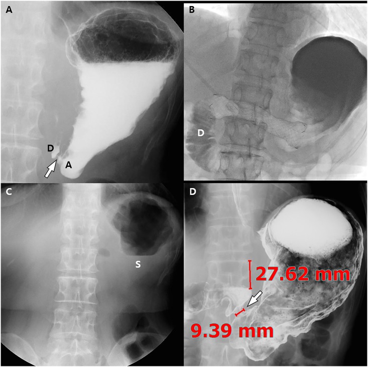
Fig 3. Images of a 68-year-old woman with impaired pyloric function after laparoscopy-assisted pylorus-preserving gastrectomy (PPG).
She underwent balloon dilatation with a 22 mm x 4 cm long balloon 3 times for 3 minutes with a 1-minute interval 19 days after PPG. (A) Post-ballooning upper gastrointestinal series (UGIS) shows tight stenosis of the pyloric canal (arrow) due to recoil of the stenosis. The pyloric canal-to-height of vertebral body ratio was 3.7. (B) Stent insertion was done for pyloric spasms with a 20 mm x 10 cm long fully covered retrievable stent. Fluoroscopic image shows good passage of the contrast agent to the duodenum (D). Stent removal was successfully done 2 weeks after stent insertion. (C) On the scout UGIS obtained 3 months after stent removal, approximately 1/4~1/2 of the stomach (S) is filled with residual food material, resulting in a semi-quantitative score for residual food of 3. (D) UGIS shows a dilated pyloric canal (arrow) and the width of the pyloric canal and the height of the adjacent vertebral body are 9.39 and 27.62, respectively. Therefore, the pyloric canal-to-height of vertebral body ratio was markedly increased from 3.7 to 34.0. Finally, her subjective symptom score for post-prandial discomfort was also markedly resolved from 10 to 3 after the stent procedure.

