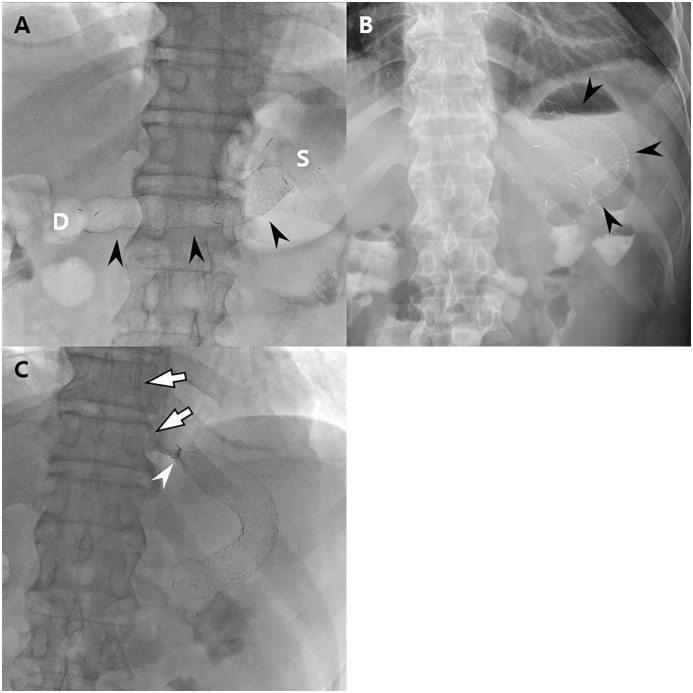
Fig 4. Images of a 56-year-old man with impaired pyloric function after laparoscopy-assisted pylorus-preserving gastrectomy (PPG).
The patient underwent balloon dilatation and subsequent stent insertion 21 and 23 days after PPG, respectively. (A) On the scout image obtained after stent insertion, the stent (arrowheads) is well placed extending from the stomach (S) to the duodenum (D) with its center located at the pyloric canal. (B) On plain abdominal radiograph obtained after 1 day, the stent (arrowheads) has migrated proximally and is located within the remnant stomach. (C) Fluoroscopic guided stent removal was done using an angiographic catheter (arrows). Note the collapsed proximal end (arrowhead) of the stent.

