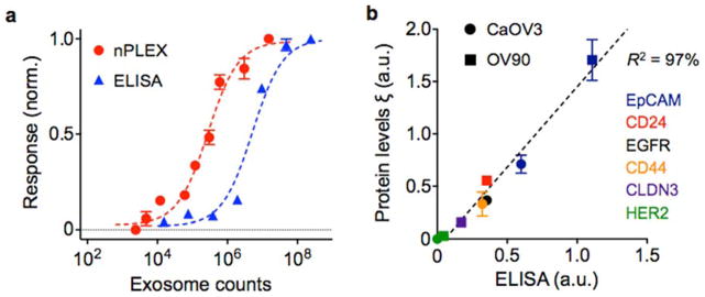
Fig. 7. Exosome quantification and protein profiling with nPLEX.
(a) Exosomes isolated from human ovarian cancer cell line (CaOV3) were introduced onto a nPLEX sensor functionalized with CD63 antibody for exosomal capture. The nPLEX platform showed considerably higher sensitivity than ELISA. (b) Comparison between nPLEX and ELISA measurements. Exosomes isolated from human ovarian cancer cell lines were used. The expression level (ξ) was determined by normalizing the marker signal with that of CD63, which accounted for variation in exosomal counts across samples. All measurements were in triplicate and the data is displayed as mean ± s.d. Reproduced from Im H, Shao H, Park YI et al. Label-free detection and molecular profiling of exosomes with a nano-plasmonic sensor. Nat Biotechnol 2014;32:490–495 with permission from Nature Publishing Group, copyright 2014 [18].

