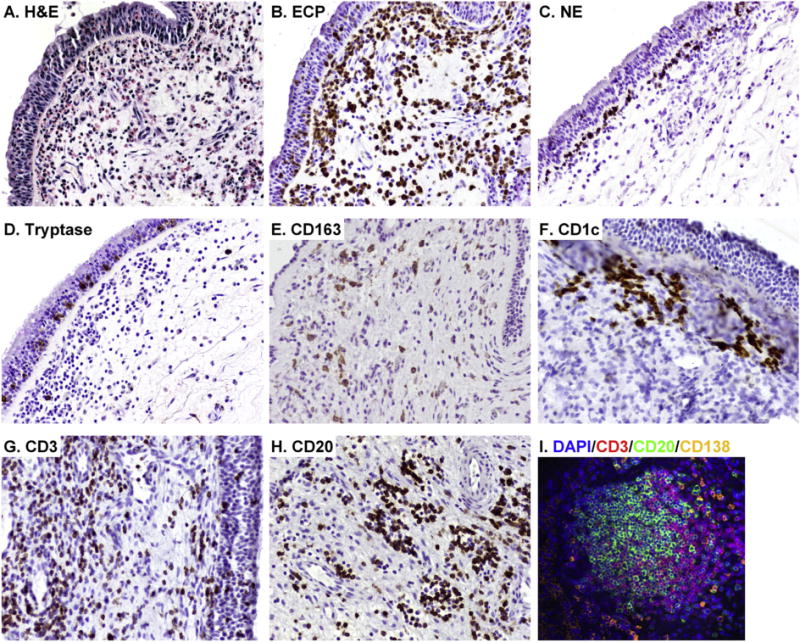
Fig. 2.

Increased presence of immune cells in NPs. Representative H&E staining in NPs is shown in A. Representative immunostainings were performed with anti-ECP mAb (EG2) for eosinophils (B), anti-neutrophil elastase (NE) mAb for neutrophils (C), anti-tryptase mAb for mast cells (D), anti-CD163 mAb for M2 macrophages (E), anti-CD1c mAb for mDC1s (F), anti-CD3 mAb for T cells (G) and anti-CD20 mAb for B cells (H). Immunofluorescence assay was performed with anti-CD3 mAb (red fluorescence), anti-CD20 mAb (green fluorescence) and anti-CD138 mAb (orange fluorescence) for plasma cells (I), Nuclei were counterstained with 4′6-diamidino-2- phenylindole (DAPI; blue fluorescence). Magnification; ×200.
