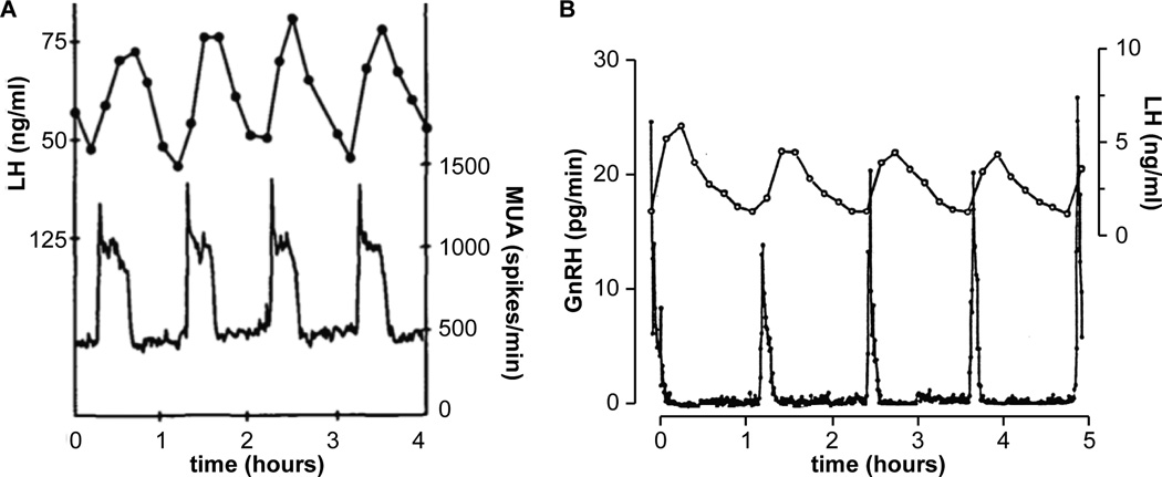
Figure 2.
Two methods for direct monitoring of reproductive neuroendocrine activity. Left, MUA measurements in the in the infundibular region of an ovariectomized rhesus monkey; peaks in MUA directly precede LH pulses. Adapted with permission from [28]. Right, simultaneous sampling of pituitary portal blood from an ovariectomized sheep at 30-sec intervals and jugular blood at 10-min intervals demonstrated a similar correlation between GnRH pulses and LH pulses. Adapted with permission from [18]. These figures have been scaled so that the X-axis is the same to facilitate comparison.

