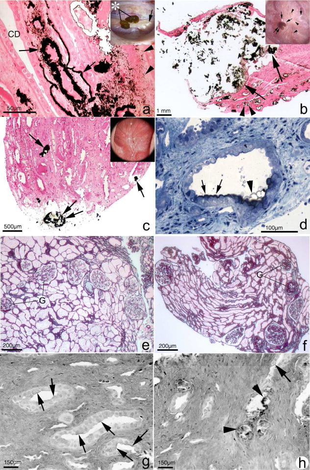
Figure 1.

Histopathology of renal papilla and cortex from CaOx stone formers. Papillary tissue of ICSF patients (panel A) shows initial sites of individual calcium phosphate deposits (arrowheads) by Yasue stain (brown-black deposits) in the basement membranes of thin loops of Henle when viewed by light microscopy. These individual deposits coalesce in the interstitial space to form islands of mineral encased in matrix material (arrows) that can extend to the basal side of the urothelium. No evidence of cell injury or inflammation has been noted in the collecting ducts (CD) or interstitium of these papilla. Endoscopic examination of the papilla of ICSF patients reveals small stones (insert panel A, asterisk) attached to the papilla by way of white or interstitial plaque (arrow). In addition to sites of interstitial plaque (panel B, double arrowhead), the papilla of patients with small bowel resection show intraluminal plugs of varying size in the IMCD (arrows). These deposits contained a mixture of CaOx, apatite and ammonium acid urate. Note the extensive interstitial fibrosis surrounding the IMCD plug. These patients also have attached stones (insert panel B, double arrow) at sites of white plaque (arrows). Areas of yellow plaque are also seen (arrowheads). Papilla of intestinal bypass patients for obesity possesses little to no interstitial plaque (panel C) but have a few intraluminal plugs in IMCD (arrow) and ducts of Bellini (double arrow) composed of apatite and CaOx. The insert in panel C shows a dilated opening to a BD (arrowhead). The initial layer of mineral to coat the apical surface of the lining cells of IMCD of these patients is Yasue positive (panel D, arrows) and particularly clear at the arrowhead. Panel E shows a cortical biopsy from an ICSF patient revealing only minimal interstitial fibrosis and glomerular (G) changes. A moderate level of change is seen panel F from a patient with small bowel resection. Panels G and H show sites of hyaluronan staining (arrows) in papillary tissue from intestinal bypass patients revealing areas of cell surface changes at sites of crystal deposits (panel H, arrowheads) or away from deposits (panel G).
